Skeletal muscle tissue: what is it and how does it work?

|
This brief text describes the striated skeletal muscle, i.e., the muscle that permits movement! |
There are different types of muscle tissue. In the widest meaning of the term, the muscle tissue allows the voluntary and involuntary movements of the organs of the body. The property of the muscle tissue involved in this task is its ability to contract, that is, to shorten its length.
The striated skeletal muscle will be described here, while the smooth and cardiac muscle types will not be considered.
 To imagine the organization of a skeletal muscle we can use the image of a pork loin. Indeed, meat is just a muscle tissue and the structure of any piece of meat is not different from that of human muscle tissue.
The base element is the muscle fibre, which is a unique cell with many nuclei resulting from the fusion of various single cells named polynuclear syncytium. Its thickness ranges between 10 and 100 μm but it can reach up to 50 cm in length. Because it is the base element, it is the sum of the contractions of the individual fibres that permits movement.
The contraction of the fibres is made possible by the repeated shortening of the single sarcomeres it contains. Every single sarcomere contains contractile proteins (myosin and actin among others) whose interaction is responsible for the shortening of the fibres and force generation. Non contractile proteins play an important support role for contractile proteins and for maintaining the structure of the fibres and of the whole muscle.
Coming back to our piece of meat, we can observe that it contains a number of groups of fibres surrounded by veins of clearer tissue. This tissue is called interstitial connective tissue and has important structural and support functions, as mentioned before. There are three types of interstitial connective tissue: the epimysium, that is a layer that encloses the whole muscle and separates it from the surrounding structures; It can be observed on the surface of the pork loin. It is a rather dense tissue resistant to lengthening. Enclosing each fascicle of fibres is the perimysium, that has similar characteristics as the epimysium. It also delimits the path of the blood vessels and of the neuronal structures in the muscle. It is visible if we observe a cross section of a piece of meat.
- Sarcomere;
- A series of sarcomeres form the muscle fibre. Each individual fibre is enclosed in the endomysium;
- Groups of fibres are enclosed in muscle fascicles by the perimysium;
- The epimysium groups all the fascicles and arranges them to form the muscle belly
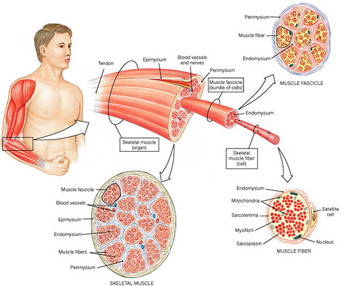
The neural impulse from the brain is transformed into contraction of the muscle fibres by a series of cell reactions induced by the neuromuscular junctions.
The skeletal muscle can undergo three different types of contraction:
- Isometric contraction: The muscle generates force without changing its length. The force produced by the muscle is equal to the external force and therefore no movement occurs. An example of isometric exercise is the bench press, in which a same position should be kept for a given period of time.
- Concentric contraction: the muscle generates force while shortening and, winning the external force it produces movement. A concentric contraction occurs when the quadriceps muscle extends the knee.
- Eccentric contraction: the muscle generates force while trying to oppose a dominant external force. The muscle slows down the movement imparted to the joint by a higher external force. An example of eccentric contraction is the action of the biceps brachii muscle while placing a glass on a table: in this case this muscle will oppose the force that gravity generates on the forearm.
- If the motor neuron is small the fibres it innervates will be able to maintain a low level of force generation over a long period of time. These fibres are called “slow twitch fibres”, and play an important role in holding a position for a long time..
- If the motor neuron is large the fibres will be able to produce force peaks in a very short period of time because they fatigue quickly. They are called “fast twitch fibres” and are responsible for explosive contractions.
A wide range of intermediate profiles exists between the two types described above.
In general, as a rule, during a movement slow twitch fibres are involved first to control the segments engaged in low intensity, durable contractions, and only at a later stage fast twitch fibres are engaged to produce increased force for a short time.
A most important property of the neuromuscular system is plasticity: in fact, it is able to adapt to the applied external loads and environmental demands. If a muscle is stimulated correctly it reacts by increasing its force and trophism. Muscle hypertrophy results from the increase of protein synthesis inside the individual fibres, that increase in size but not in number. Instead, the adjustments of the nervous system are responsible for the increased force of a muscle. Among them, neural disinhibition and an increased activity of the cerebral cortex are worth to be mentioned, as well as the higher degree of inhibition of antagonist muscles, which allows agonist muscles to generate more force.
Conversely, inactivity even for a few weeks causes a considerable reduction of muscle force and size. Proportionally, this degeneration affects slow twitch fibres more markedly.


Agonist muscles are primarily responsible for the start and execution of a movement. For instance, the hamstrings are the agonist muscles in the flexion of the knee and extension of the thigh.
Antagonist muscles oppose the movement of the agonist muscles. Referring to the hamstrings, the quadriceps muscle can be defined as their antagonist.
Synergist muscles are involved to a limited extent in performing a movement. Each movement requires the involvement of many synergist muscles.
Muscles can be monoarticular or biarticular. Monoarticular muscles act on only one joint, i.e., they perform only one movement on only one segment of the body (for instance, the piriformis muscle acts only on the hip joint). Biarticular muscles are much more complex and act on two adjacent joints (the rectus femoris muscle extends the knee and flexes the hip). This distinction is important mainly if one considers that biarticular muscles are much more prone to injury.


